Modeling the Optics of the Eye
Manushi Shah
Mentor: John Noé
Laser Teaching Center
Department of Physics & Astronomy
Stony Brook University
Introduction
During the beginning of the year, Dr Noé used to tell everyone that we should read more about the topics that we are interested in. I used to search about the laser eye surgery. I was so sure that I will do my project on that, but I didn't realize how different, but interesting, my project became. While I was learning more about the eyes, I found learning about the eye defects more interesting. I was also interested in learning more about the different kinds of aberrations in my eyes. Since I have cylindrical numbers, I wanted to understand that more deeply. So my project starts out with modeling the simple eye and its different components and what each does. As seen in my journal, I modeled simple eye, approximating it as a water ball. It was my first step to learn more about the BEAM 2 program that I later found out to be so helpful in producing exact ray diagrams.
Background
By searching online, I found more information about the necessary components that are responsible for the refraction of the incoming rays. The optical components of a normal eye are the cornea, the aqueous humor, the crystalline lens, and the vitreous humor. About 2/3 of the total refraction occurs in the cornea, mostly at the front or the air-cornea boundary. I also found out that remaining 1/3 of the refraction occurs in the crystalline lens. The total power of the crystalline lens is the sum of the contributions from the front and back surfaces; the back surface is more curved and contributes more than the front. The corneal power is about 43 D. As we know, the normal or relaxed eye brings light rays from far away (infinity) to a sharp focus on the retina. To view a near object, the ciliary muscles increase the curvature of the back surface of the lens thereby increasing its focal power. The relaxed lens power is about 19 D. The total eye power is about 62 D. We were able to estimate this by approximating the eye length to be about 25 mm and then taking the inverse of its focal length.
The BEAM2 Program
Learning about the program wouldn't have been so hard if we had the manual in the first place. But it was still fun using the trial and error method to produce the different diagrams. The BEAM2 program is an inexpensive and easy to use tool for exact tracing of light rays through the boundaries between media or at reflective surfaces. It is limited to spherical and conical surfaces of revolution about the z-axis. Surfaces are described in an Optics Table formatted in plain text. Parameters that can be specified include: shapes, indexes, curvature, diameter and position of the lens, mirrors or other surfaces. Light rays are described in a separate Ray Table by their initial position and inclination with respect to the x and y axes. The most interesting part was looking at the final image after making many changes. BEAM2 results appear as on-screen diagrams that can be transferred to the other programs for further editing and printing. The diagrams can be shaded to indicate areas of different index of refraction.
Description of the Table
Below is the simple table that I used to produce the normal eye. We decided to use one
book and use their approximated values for the indices of refraction of different surfaces and
thicknesses of the lens and cornea. The following describes the meaning of the columns
- Index
- The indices of refractive surfaces that I used in the diagram. The indices are defined for the surfaces to the left of the described surface because the column is on the left side.
- Zvx
- Vertex position of the surface. I used a random position for the first surface (cornea), but for the next surface position I used the corneal thickness (.6 mm) to find out the eyeball position.
- Curv
- This column defines the curvature of each surface. I calculated the curvature by finding the lens' radius of curvature and then taking its' inverse. First, I really tried to approximate that becuase I wasn't sure how to calculate it. Later, Dr. Noe told me how to calculate it using the lens maker's formula and it became so easy.
- Mir/Lens
- Defines whether the surface is a lens, screen, mirror or iris.
- Diameter
- diameter of the element.
- Shape
- Shape of the surface. 1 defines the spherical shape.
Optical Table of the Model Eye
| 7 surfaces | |||||
|---|---|---|---|---|---|
| Index | Z vx | Curv | Mir/Lens | Diameter | Shape |
| 1.00 | 1.4 | .1144 | Lens | 12 | 1 |
| 1.3762 | 2 | .09091 | Lens | 22 | 1 |
| 1.376 | 6.5 | .083 | Lens | 8.5 | 1 |
| 1.47 " | 10.3 | -.222 | Lens | 8.5 | 1 |
| 1.336 | 24 | -.09091 | Lens | 22 | 1 |
| 1.336 | 24 | -.09091 | Lens | 4 | 1 |
| 1.00 | 24.05 | -.09091 | Screen | 4 | 1 |
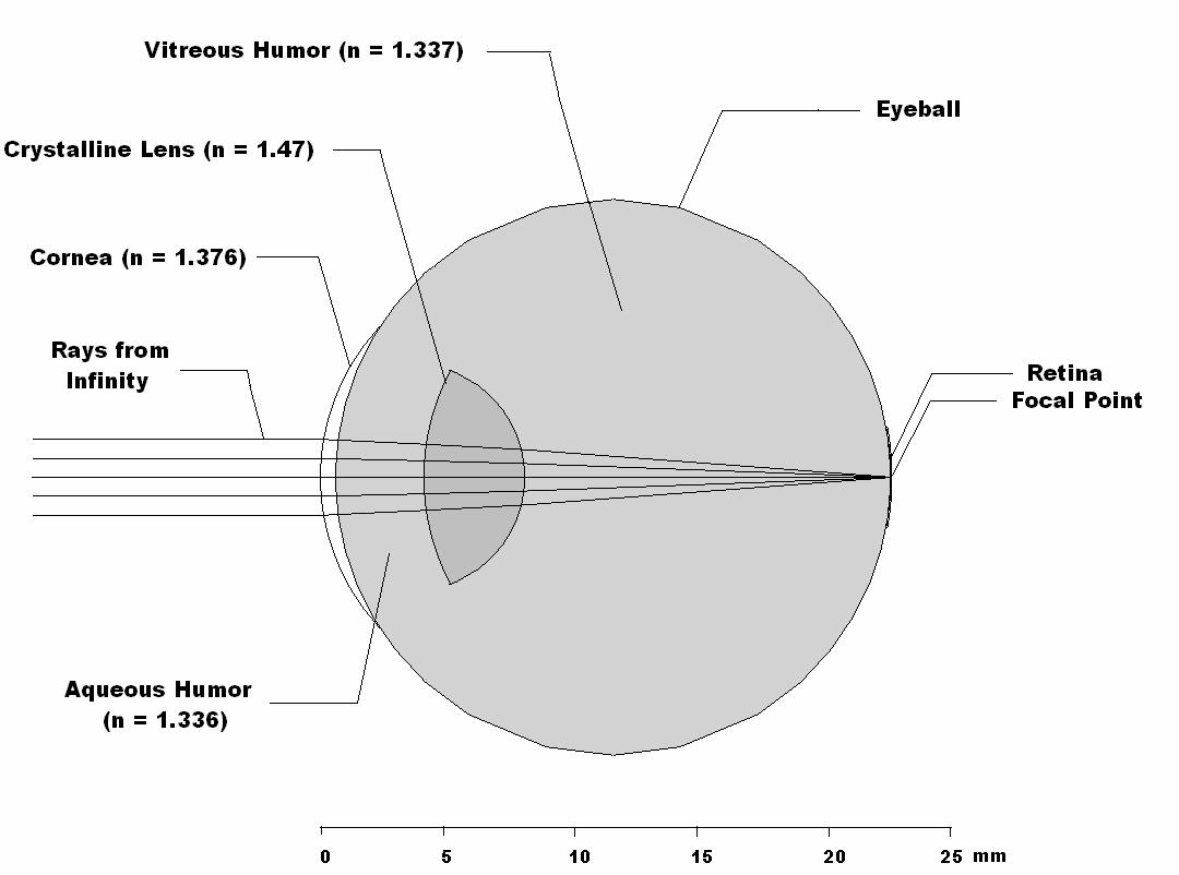
This is the model of a normal eye made with the BEAM2 program. I used following approximated values from the literature: eyeball diameter = 22 mm, cornea diameter = 12 mm, cornea thickness on axis = 0.6 mm, distance from cornea to lens = 4.5 mm, lens thickness = 3.8 mm. With these dimensions the focus point was exactly on the retina.
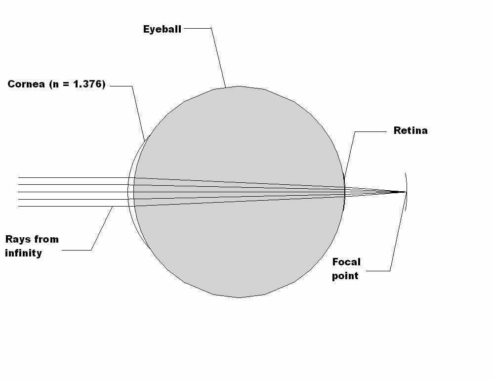
This is a hypothetical model of an eye without a crystalline lens. Here the focus point is far-behind the retina. This shows that about 2/3 of the eye power comes from the cornea.
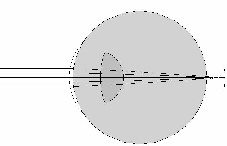
Here is the model of a hyperopic eye (far-sightedness). The rays come to a focus behind the retina. Here one of the limitations that Dr Noe later pointed out is that the rays actually don't come from the infinity as the image shows, but from the near point that I can't show because the object distance is much greater than the eyeball size. The shortness of the eyeball or an inability of the lens to become round causes rays to focus behind the retina.
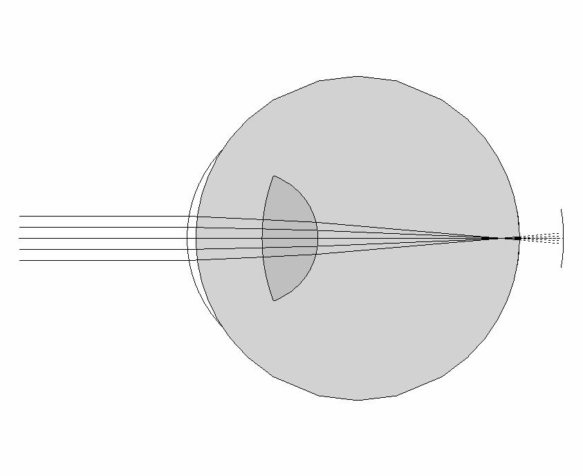
This is the model of a myopic eye (near-sightedness). The rays come to a focus in the vitreous humor inside the eye rather than on the retina. Steepness of the cornea or increased size of an eyeball can cause this condition.
Lens Calculations
Lens Maker's Formula
I used the Lens Maker's Formula to calculate the radii of curvature of the cornea and the
crystalline lens. The lens power P for each spherical boundary between two media is given by
P = [(n_lens -– no)/(_o)] (1/R)
where n_lens and n_o are the indices of refraction of the two media and R
is the radius of curvature of the spherical boundary between them, in meters.
The cornea has a greater focusing power than the crystalline
lens because the index of refraction change from air (n = 1) to cornea is
much greater than that between the surrounding media and the crystalline
lens.
Gullstrand's Equation
I used the Gullstrand's Equation to calculate the
effective power of a thick lens P from the powers of
its two surfaces p_1 and p_2. P = p1 + p2 – p1 * p2 * (d/n)
Here d is
the distance on axis between the two surfaces and n is the index of
refraction of the material between them.
This equation allowed me to
calculate the overall effective power of two surfaces in a thick lens, or
two thin lenses separated by some distance, for example an eyeglass lens and the eye itself.
Facts about Eyes
I have here some of the most interesting facts. I always used to think
that there is such a thing as the perfect vision. But I found out that
no one has a perfect vision, not even those with "20/20" vision. The
Snellen eye chart determines the ratio of the normal vision to the
patient's vision. The normal eye with 20/20 vision is called an
emmetropic eye.
The focusing power is measured in diopters. A diopter is the
reciprocal of the focal length measured in meters. So our eye glasses
are measured in few diopters.
Myopia usually occurs in younger children. Children can clearly see near objects yet are unable
to focus on distant objects. Hyperopia, a common visual defect in
adults, is a condition where one is unable to see nearby objects yet
is able to focus on objects that are farther away. Older people lose
the ability to change the focus of the eye(accommodate). This
condition is called presbyopia. It usually starts at about the age 42.
More than 98% of our world is affected by aberrations, deviation
of rays from the expected path for an ideal lens.
I even found
out that coma, one of the aberrations, causes a variation in
magnification over the plane. Astigmatism is another type of
aberration that occurs due to the difference in the focusing of the
rays in perpendicular planes.
Spherical, another type of aberration, causes image imperfections due to increased refraction of
light.
Different objective tests to measure the visual acuity include direct ophthalmoscope, keratometry and
retinoscope.
References
1. Sharma, K. "Principles and Application" publication: Academic
Press. Copyright: 2006.
2. Nave, R. "Design of Scale-Model
Eye" http://hyperphysics.phy-astr.gsu.edu/Hbase/geoopt/gullcal.html#c1
3. Bass, Michael. "Handbook of
Optics" publication: McGraw-Hill. Copyright: 2001. Edition: 2nd. Volume: 3.
4. Arell, Antti. "Experiments on a Model Eye." American Journal of
Physics.http://scitation.aip.org/ajp/
5. Gregory, P. "Eye Models" http://science.tjc.edu/images/eye&ear/eye_models.htm
6. Howell, William. "The Sense of Vision." An American Text-book of
Physiology.
[ Eye Model]
Manushi Shah