
Background
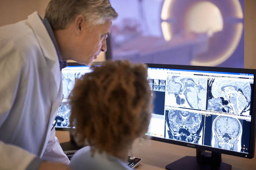
The advent of medical imaging of the anatomy's structure and function has allowed radiologists to view a patient's anatomy without the immediate need for invasive surgery. Transmission Tomography (TT), such as Computed Tomography (CT), allows the radiologist to view the patient's anatomical structure, while Emission Tomography (ET) allows the radiologist to view the patient's anatomical function. Positron Emission Tomography (PET) and Single Photon Emission Computed Tomography (SPECT) are common techniques for imaging anatomical function. A common algorithm used in SPECT image reconstruction is the iterative maximum likelihood expectation maximization (ML-EM) reconstruction algorithm with the ordered-subsets (OS) strategy, the combination of which is commonly referred to as the OS-EM algorithm. Although the OS-EM algorithm achieves a good quantitative reconstruction, there are limitations in clinical use. One drawback is the high computational cost of the algorithm resulting from the large vectors and matrices associated with producing a high-resolution image reconstruction. Research efforts have been devoted to mitigate drawbacks by (1) developing efficient simulators for the re-projection and back-projection cycle, such as by the use of the geometry warping with distance-dependent convolution or the recursive ray-tracing with geometry symmetries; and (2) investigating sophisticated strategies to speed up the convergence to a satisfactory result, such as the OS technique. Although a significant speed gain was observed by the addition of the OS technique, the reconstruction time is still typically too long for acceptable clinical use. Improved techniques for image reconstruction for SPECT images are desired.
Technology
Reconstruction of high resolution medical images is clinically desired, especially in dynamical studies. Researchers at Stony Brook University have developed innovative software models to model the data collection and processing for ultra fast image reconstruction. By integrating these models into the GPU hardware architecture, ultra fast image reconstruction has been realized. This speedup can be applied to image segmentation and image rendering.
Application
- Reconstruction of high resolution medical images - Image segmentation and image rendering
Inventors
Jerome Liang, Professor, Radiology
Zigang Wang, Research Associate, Radiology
Licensing Potential
Licensing
Licensing Status
Available for licensing.
Licensing Contact
Donna Tumminello, Assistant Director, Intellectual Property Partners, donna.tumminello@stonybrook.edu, 6316324163
Patent Status
Patented
8687869
Tech Id
7861